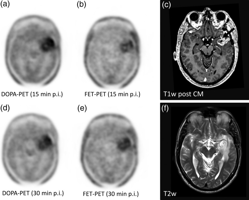
Fig. 2.
In a participant with glioblastoma, the typical contrast media enhancement in T1-weighted MRI for high grade gliomas was found (c, arrow), surrounded by edema with higher signal intensities in T2w (f). In DOPA-PET, a washout from the 15-minute (a) to the 30-minute (d) p.i. frame and a moderate washout in FET-PET(b) versus (e) were observed.

