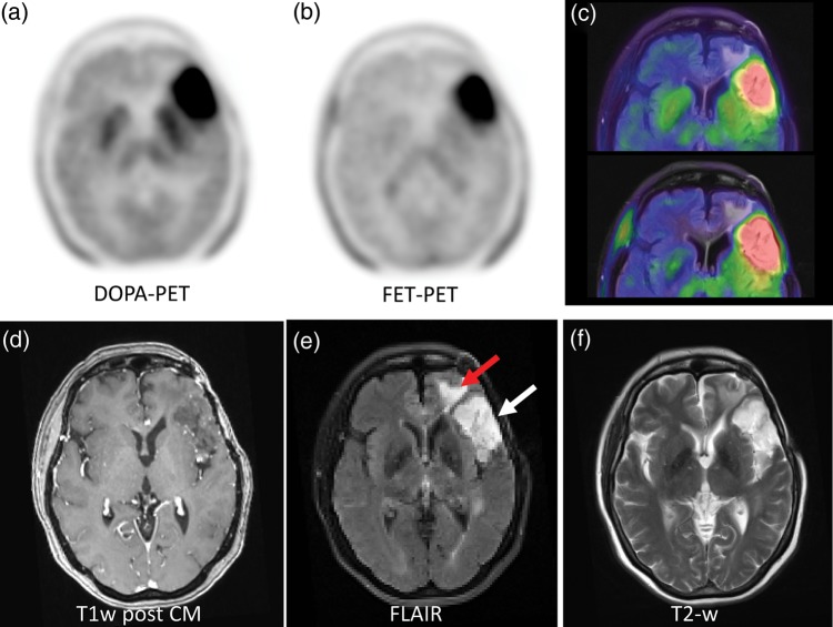
Fig. 4.
A participant with low-grade glioma, which is rarely delineable in contrast-enhanced T1w (d). The FLAIR (e) and T2 (f) weighted sequences demonstrate elevated signal intensity in the anterior temporal (e, white arrow) and the lateral frontal lobe (e, red arrow) but cannot differentiate between tumor and edema. With both, DOPA (a) and FET (b) PET, the malignant tissue was equally demarcated as presented in the fusion images (c).

