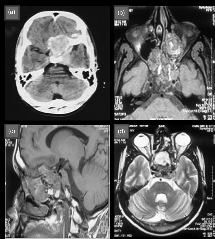
Fig. 1.
(a) Contrast-enhanced computed tomographic image showing suprasellar space-occupying lesion with heterogenous contrast enhancement. (b) and (c) T1 weighted magnetic resonance image showing iso to hypointense lesion in suprasellar region with foci of high-signal intensity and extension to left orbit and ethmoid sinus. (d) T2 weighted magnetic resonance image showing hyperintense lesion in suprasellar region.

