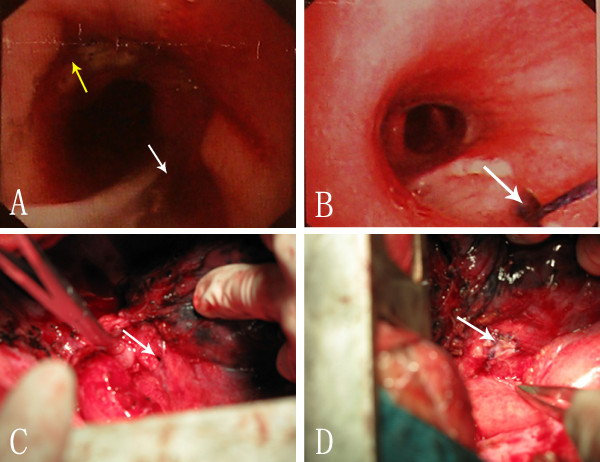
Figure 1.
Preoperative endoscopy and intraoperative view of patient 1. (A) Esophagogastroscopy showing the anastomotic stoma (yellow arrow) and a fistula between anastomotic stoma and right intermediate bronchus (white arrow). (B) Bronchoscopy showing fistula (arrow head) within the right intermediate bronchus surrounded by large mucosa erosion. (C) Intraoperative view of an oval fistula (arrow head) in the right intermediate bronchus after resection the diseased gastric tube. (D) Intraoperative view of the repair of right intermediate bronchial defect with a pedicled pericardial flap using interrupted suture (arrow).

