
Figure 2.
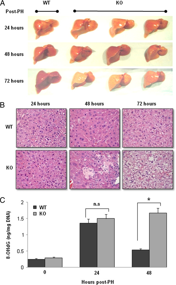
Oxidative DNA damage in the remaining liver after PH. (A) The gross morphological changes after 70% PH. There is no difference between WT and IL-6 KO mice on day 1 and day 2 after PH. However, on day 3 after PH, IL-6 KO livers show pale-yellowish color and the liver size did not increase. In contrast, the WT liver increased in size and had a normal color. Representative photographs of n = 3 individual samples per group. (B) H&E staining demonstrated that necrosis appeared in liver tissue sections recovered from IL-6 KO mice at 48 and 72 hours after PH in IL-6 KO mice. No necrosis/apoptosis appeared in liver sections from WT mice after PH. (C) Levels of 8-OHdG in DNA of the remaining liver after PH. Data represent mean ± SEM (n = 3). n.s., no significant difference. *P < 0.05.