Fig 2.

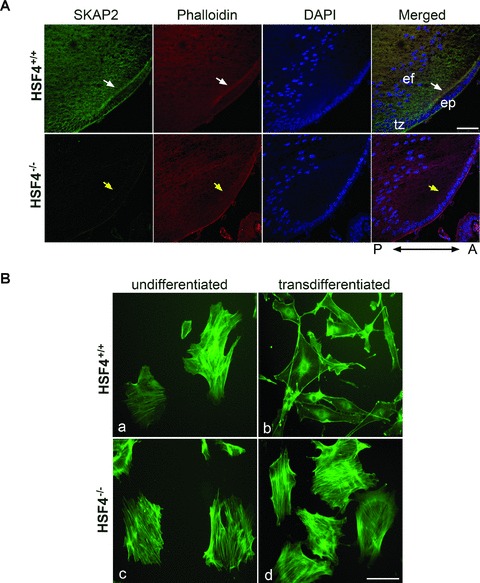
Defective reorganization of the actin cytoskeleton in HSF4−/− mouse lens cells. (A) SKAP2 was colocalized with F-actin at the anterior tip of elongating fibre cells in HSF4+/+ lens (white arrows), but not in HSF4−/− lens (yellow arrows). Mid-sagittal lens sections that were prepared from wild-type or HSF4−/– mice at postnatal day 4 were stained with phalloidin-Alexa Fluor 555. The images were taken under a confocal microscope. The bar represents 50 μm. ep: epithelium; tz: transition zone; ef: elongating fibre cell; A: anterior; P: posterior. (B) The HSF4−/− cells failed to reorganize their actin cytoskeleton in vitro. The primary undifferentiated lens cells (parts a, c) were cultured in medium with serum. To induce transdifferentiation in vitro, the primary lens cells were starved for 24 hrs and subsequently treated with 40 ng/ml of FGF-b for 36 hrs (parts b, d). The cells were fixed and stained with phalloidin-FITC. Bar 100 μm.
