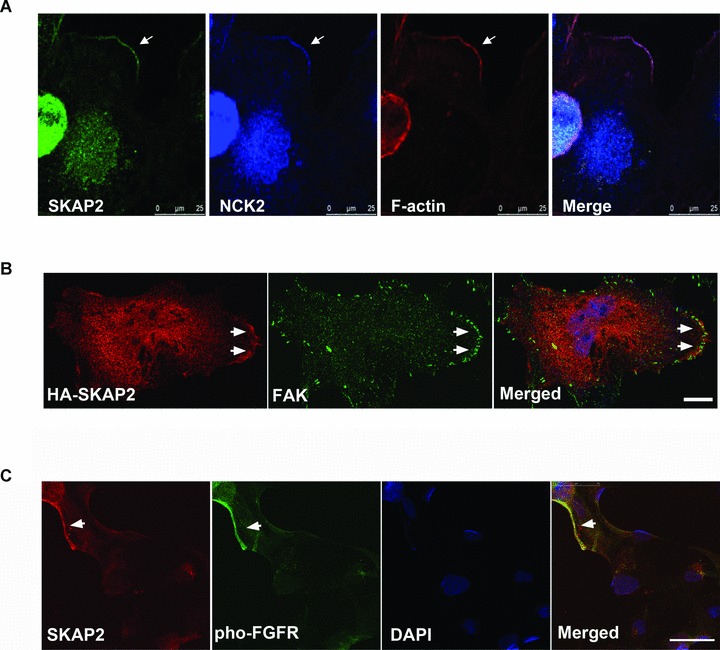
Fig 6.

SKAP2 accumulates with NCK2, FAK and FGF receptor at the leading edges. (A) Endogenous SKAP2, NCK2 and the cortical actin fibres colocalized at the leading edge of the lamellipodia. The SRA01/04 cells were stained with rabbit anti-SKAP2 antibody, monoclonal mouse anti-NCK antibody and Phalloidin Alexa Flour 555. The arrow indicates the lamellipodia. Bar 25 μm. (B) The cells expressing HA-SKAP2 were stained with mouse anti-HA and rabbit anti-FAK antibodies. The accumulation of SKAP2 and FAK at the leading edge is indicated by arrows. Bar 10 μm. (C) Colocalization of SKAP2 and phospho-FGFR in the SRA01/04 cells. The cells were starved and treated with FGF-b. Endogenous SKAP2 and the phospho-FGF receptor (Tyr653/654) were visualized with rabbit anti-SKAP2 antibody and monoclonal mouse anti-Phospho-FGFR antibody. Bar 50 μm.
