
Fig 2.
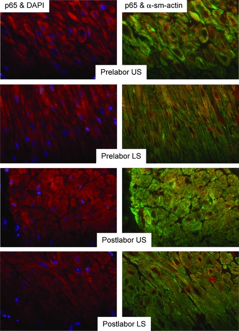
No labour-associated or regional differences in expression or nuclear localization of p65 in myometrium at term. Paraffin-embedded tissue taken from matched upper and lower-segment myometrial biopsies collected prior to (L−) or following (L+) labour onset was subject to immunofluorescent analysis, using p65 with red-fluorescent Alexa Fluor 594 dye and a-smooth muscle actin (α-sm-actin) was visualized with green-fluorescent Alexa Fluor 488 dye. Cell nuclei were visualized with DAPI (blue). (L−, n=5, L+, n=6). Objective lens used was x40.