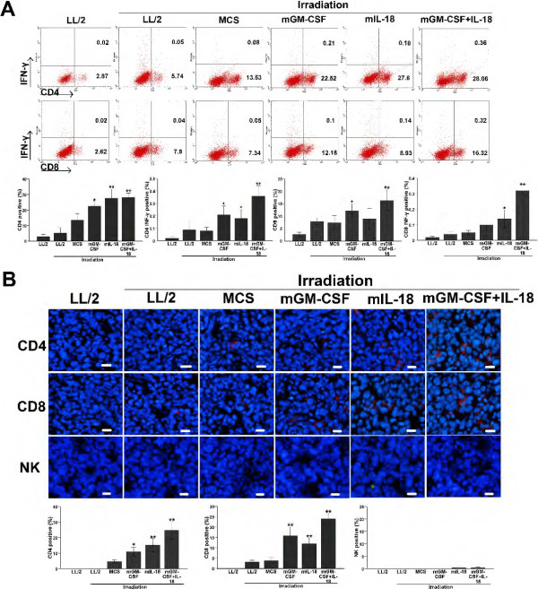
Figure 5.
Increased proliferation of CD4+INF-γ+T, CD8+INF-γ+ T in spleen and infiltration of CD4+T, CD8+T in tumors. Spleen lymphocytes were isolated and stained for CD4, CD8 and INF-γ double staining antibodies by flow cytometry; Tumor tissue was obtained 3 days after the last measurement of tumor volume, frozen sections were used for analysis of CD4, CD8 T and NK cell infiltration. (A) The proportion of CD4+INF-γ+ T, CD8+ INF-γ+ T in co-expression IL-18 and GM-CSF-treated mice was significantly higher than control groups (P < 0.01, n = 7). Experiments were performed in triplicate and repeated three times. (B) Immunofluorescence staining of tumor tissue with CD4, CD8 and NK antibody showed that CD4+, CD8+ T cell infiltrations was significant enhanced in co-expression IL-18 and GM-CSF-treated group as compared with control groups (P < 0.01, n = 7) (original magnification, ×200).

