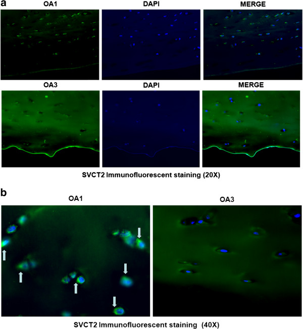
Figure 2.

Representative immunofluorescence localization of SVCT2 protein in OA1 and OA3 Collins scale grade human cartilage tissue. a) 20× and b) 40×. The immunofluorescence analysis was performed using a polyclonal antibody specific for SVCT2. Secondary antibody was anti-goat IgG labeled with FITC. The nuclei were stained with Hoechst dye.
