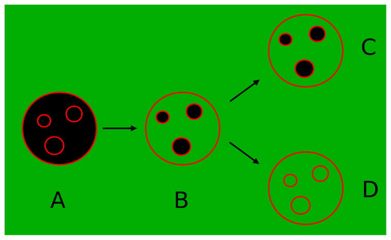
Figure 1.
The concept of the experiment. GUVs prepared with inner vesicles are added to a solution containing peptide and a water-soluble fluorophore (green). The membrane of the vesicles is shown in red. Initially (A), the interior of the vesicles has no fluorescence (black). In (B) the peptide induced flux into the outer vesicle. If the peptide does not translocate across the outer membrane, the inner vesicles remain dark (C). Appearance of fluorescence inside inner vesicles indicates that the peptide translocated across the membrane of the outer vesicle (D).

