
Figure 3.
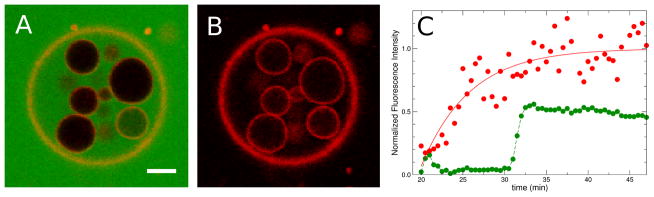
Large GUV with inner vesicles 44 min after addition of the peptide Rh-TP10W. (A) CF and Rh channels. (B) Rh channel only. Scale bar, 10 μm. (C) Fluorescence of the inner vesicle at the bottom right in A and B, as a function of time. Red points, Rh channel, showing Rh-TP10W on the inner vesicle membrane. Green points, CF channel, showing flux into the inner vesicle (intensity relative to the outer membrane). The red line is 1 − exp[−(t − to)/τR] where τR = 5.8 min and to ≈ 20 min (beginning of recording). The green line is only to guide the eye.