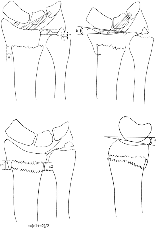
Fig. 1.

Illustration of radiographic parameters on radial translation (a), radial inclination (b), radial shortening, which was calculated as average shortening of radial shaft and ulnar shaft (c), ulnar variance (d), and displacement of the ulnar styloid fragment (e) on PA view and volar tilt (f) on lateral view.
