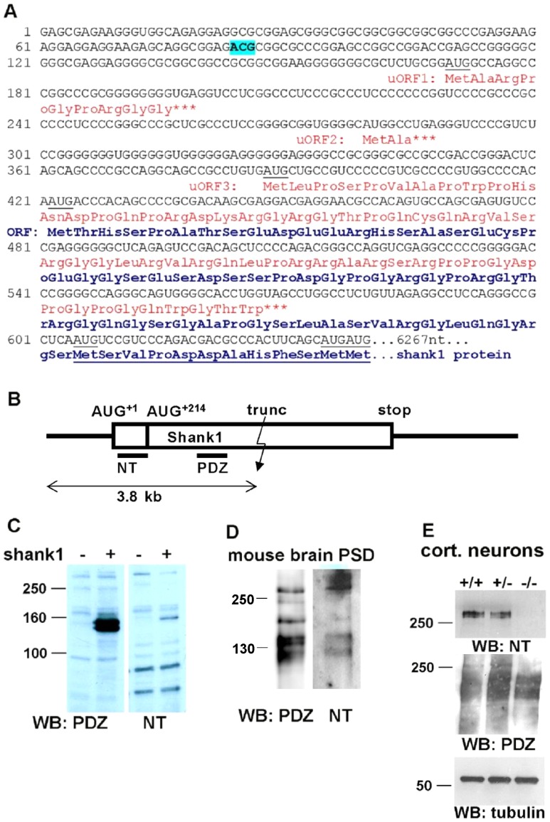
Figure 1. Alternative translational start sites in the Shank1 5′UTR.
A. Sequence of the 5′ region of the human Shank1 mRNA; seven possible AUG start codons are underlined. Translation initiation at the first three of these leads to premature termination of translation (uORFs 1–3; translation products labeled in red). AUG+1, and a cluster of three closely appositioned AUGs (collectively termed AUG+214) may both initiate translation of the full-length Shank1 ORF. Start at AUG+1 leads to an N-terminal elongation of the Shank1 protein by 70 amino acids. Amino acid sequence common to both variants is underlined. An ACG codon (nt 84–86) which becomes important later in this manuscript is indicated. B. Scheme of the Shank1 mRNA; UTRs are indicate by a line, the coding region is boxed. In frame AUGs of the main ORF, the size of a truncated expression construct of 3.8 kb, and the position of different antigenic domains (NT, PDZ) used for raising antisera are indicated. C. HEK cells were transfected with no plasmid (−) or with the truncated Shank1 expression construct (+) encompassing the first 3800 bp of the Shank1 cDNA, including the complete 5′UTR. Cell lysates were analyzed by Western blotting using an antiserum directed against the PDZ domain (left), or against the alternative N-terminal region generated by initiation at AUG+1 (NT; right). Note that the PDZ antibody recognizes two bands at approximately 130–140 kDa (calculated: 116 and 123 kDa), whereas the NT antibody recognizes only the upper one of these two bands. D. A PSD preparation derived from mouse brain was analyzed by Western blotting with PDZ and NT directed antibodies. E. Cortical neurons were prepared from wt (+/+), heterozygous (+/−) and Shank1 deficient (−/−) mice. Cells were lyzed and analyzed by Western blotting using anti-Shank1-NT (upper), anti-PDZ (middle) and anti-tubulin (lower panel) antibodies.

