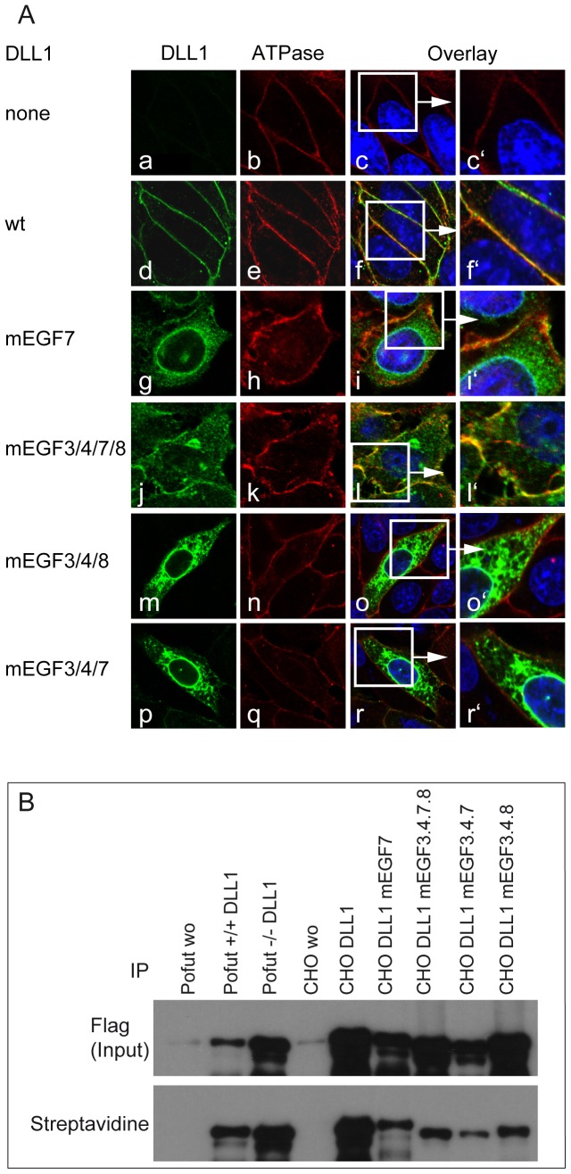
Figure 2. Subcellular localization of DLL1 protein variants in CHO cells.
(A) Confocal images stained with antibodies against DLL1 and a marker for the cell surface (Sodium potassium ATPase). (a-r) CHO cells stably transfected with control (a-c), wild type DLL1 (d-f), or mutant DLL1 (g-r) show wild type DLL1 on the surface (d-f′) and the protein variants DLL1mEGF7, DLL1mEGF3/4/7/8, DLL1mEGF3/4/7 and DLL1mEGF3/4/8 were detected on the cell surface and intracellularly (g-i′, j-l′, m-o′, p-r′). (B) Western Blot analysis of DLL1 protein variants after cell surface biotinylation, cell lysis and immunoprecipitation with Flag-Agarose beads (Flag Input) and Streptavidin-Sepharose to detect the cell surface biotinylated fraction (Streptavidin). Cells and constructs are indicated above the lanes.

