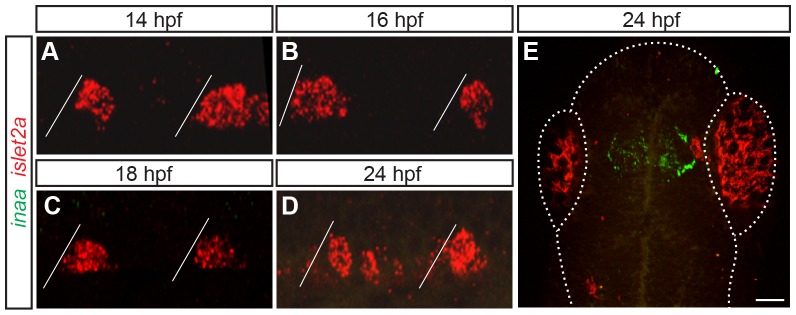
Figure 3. inaa is expressed in the zebrafish head and not in PMNs.

(A–E) Z-projections of confocal stacks of embryos labeled with inaa and islet2a riboprobes. Diagonal lines represent somite boundaries. Between 14 hpf and 24 hpf, inaa is never coexpressed with islet2a in the trunk (A–D). At 24 hpf, inaa expression can be seen in a region of the head that corresponds to the approximate location of the diencephalic ventricle (E). Boundaries of the fish head and eyes are marked with a dotted line. Scale bar 10 µm in A–D; 50 µm in E.
