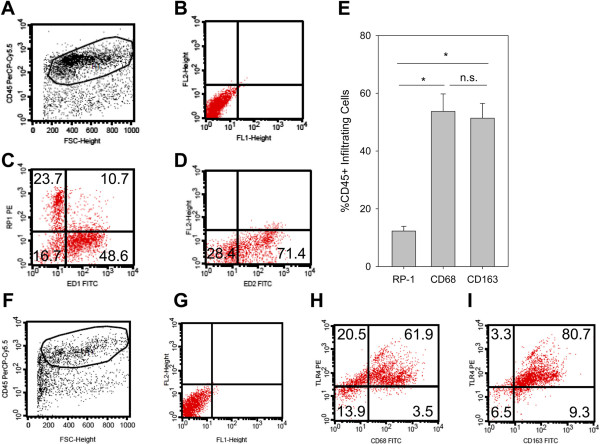
Figure 2.
Characterization of infiltrating leukocytes in the inflamed paw. Rats were injected with CFA for 4 d. (A) Cells suspensions from subcutaneous paw tissue were first stained for CD45 to gate on hematopoetic cells (x-axis: forward scatter [size], y-axis: CD45-PerCp-Cy5). (B) The dot plot (x-axis: FITC, y-axis: PE) shows unstained controls from cells gated on CD45. CD45+ cells were stained for RP-1 (neutrophils), CD68 (M1, proinflammatory macrophages) (C, x-axis: CD68-FITC, y-axis: RP-1-PE) as well as and CD163 (M2, antiinflammatory macrophages) (D, x-axis: CD163-FITC, y-axis: PE). Representative examples are shown (n = 8). (E) The percentage of infiltrating leukocyte subpopulations were measured (n = 8, ANOVA on Ranks, Student-Newman-Keuls). (F-I) TLR4-Expression in the paw was analyzed in leukocytes gated on CD45+ (F, x-axis: forward scatter, y-axis: CD45-PerCp-Cy5 fluorescence). TLR4-Expression in CD68+ M1 macrophages (H, x-axis: CD68-FITC, y-axis: TLR4-PE) and in CD163+ M2 macrophages (I, x-axis: CD163-FITC, y-axis: TLR4-PE) and compared to the unstained control (G, unstained control, x-axis: FITC fluorescence, y-axis: PE fluorescence). Representative examples are shown (n = 2).

