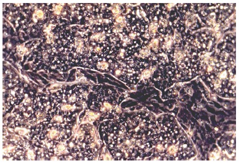
Figure 1.

Primary duck embryonic hepatocytes grown in CeLLBINDTM plates at day 3 of cultivation. Monolayers of hepatocytes, which show typical polygonal morphology, are interrupted by areas of non-parenchymal cells (light microscopy, phase contrast, x 200).
