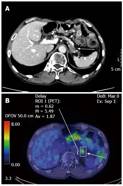
Figure 1.

Enhanced computed tomography (A) and 18F-2-fluoro-2-deoxy-D-glucose positron-emission tomography (B) showed a tumor of the pancreatic body (arrows). The major axis of the tumor was 15 mm. Maximum standardized uptake value of the lesion was 5.49.
