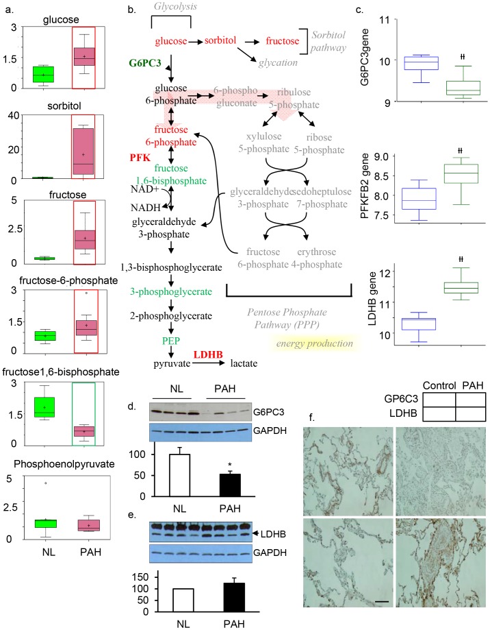
Figure 2. Glycolysis is significantly upregulated in the PAH lung.
a) In all graphs, data for normal lung are shown in green boxes and data for the PAH lung are represented in pink boxes. Quantities are in arbitrary units specific to the internal standards for each quantified metabolite and normalized to protein concentration (N = 8 for each box). PAH patient samples exhibited higher levels of glucose, sorbitol, fructose, and fructose 6-phosphate. This metabolic disruption can contribute to the formation of advanced glycolytic end products that have been shown to directly contribute to the severity of PAH. b) The classical glycolysis/pentose/energy pathways are shown. c)Three genes encoding G6PC3, PFKFB2, and LDHB were significantly changed in PAH lung compared with NL, as shown in all graph) (G6PC3 (p = 8.54e−05), PFKFB2 (p = 2.6e-04) and LDHB (p = 2.19e−09). d) Western Blot analysis of G6PC3 (n = 4/each group) and e) LDHB expression in normal and PAH lungs (n = 4/each group). Lung lysate (20 ug per lane) was loaded and immunoblotted with antibody against G6PC3 or LDHB and GAPDH (loading control). Consistent with a significant decrease of G6PC3 and an increase of LDHB gene expression in PAH, protein expression for G6PC3 (39KD) was significantly decreased while LDH (37KD) was significantly increased in PAH lungs compared with NL lungs. Densitometric analysis of G6PC3 and LDHB were normalized to the intensity of the respective GAPDH band. Data are expressed as mean ±SD (n = 4). *P<0.05 versus NL. f), Representative images of G6PC3 positive immunostaining in the collagen fibers of pulmonary vascular tissue, which was decreased in PAH. Increased LDHB positive staining was found in pulmonary vascular smooth muscle tissue in the PAH lung (bar = 1∶400).

