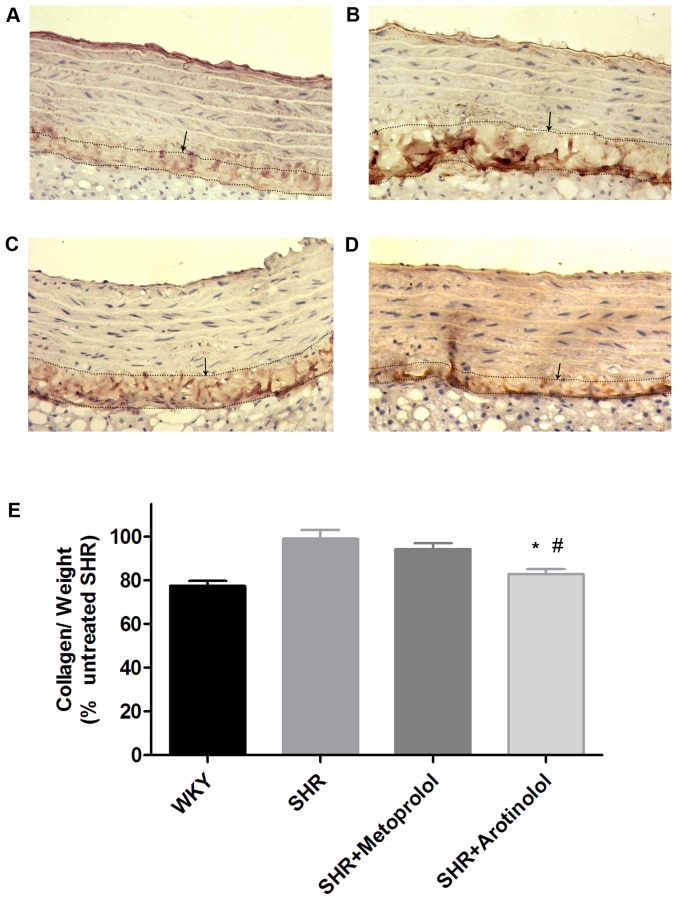
Figure 7. Collagen contents in rat aortas by immunohistochemistry and biochemistry assay.
Immunohistochemistry of collagen I, which was mainly distributed in the adventitia of rat aortas (×200). (A)WKY, (B)SHR control, (C)SHR treated with Metoprolol, (D) SHR treated with Arotinolol. (E) Summarized data showing the changes in collagen contents of aortas in WKY, SHR control, SHRs treated with metoprolol or arotinolo by chloramine T and paradimethylaminobenzaldehyde method. *P<0.05 vs. SHR control, # P<0.05 vs. SHR treated with metoprolol. n = 12 in each group.

