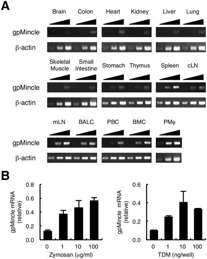
Figure 1. Expression of gpMincle mRNA.
(A) Tissue distribution of gpMincle mRNA. mRNA expression of gpMincle in indicated tissues (cLN, cutaneous lymph node; mLN, mesenteric lymph node) and cells (BALC, bronchoalveolar lavage cell; PBC, peripheral blood cell; BMC, bone marrow cell; PMø, thioglycollate-elicited peritoneal macrophage) was detected by PCR. PCR was performed by increased cycle numbers (20, 24, 28 for β-actin and 32, 36, 40 for Mincle). (B) gpMincle mRNA is induced upon stimulation. Macrophages were stimulated with indicated concentrations of zymosan (left panel) or TDM (right panel). mRNA expression of gpMincle was analyzed by RT-PCR at 8 h after stimulation. Data are presented as mean ± s.d. (B) and representative of two separate experiments.

