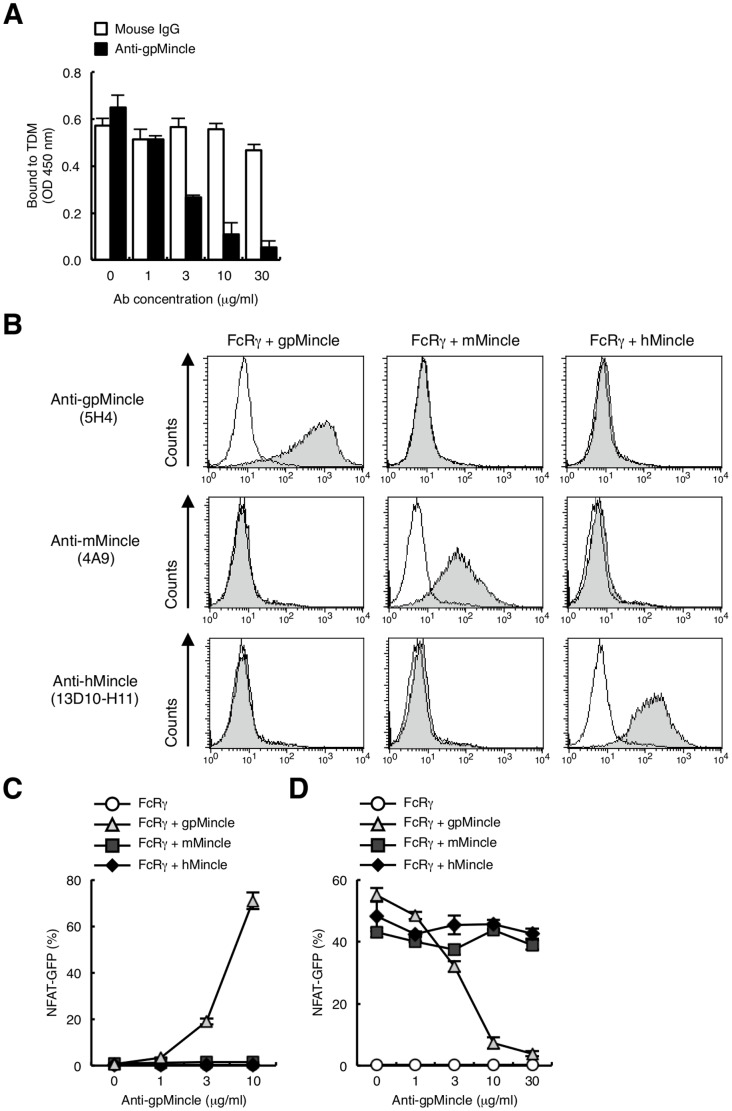
Figure 5. Establishment of anti-gpMincle mAb.
(A) Anti-gpMincle mAb blocks interaction of gpMincle-Ig with TDM. gpMincle-Ig (3 µg/ml) were incubated with plate-coated TDM in the presence of anti-gpMincle mAb or mouse IgG. Bound proteins were detected with anti-hIgG-HRP. (B) Surface staining by anti-gpMincle mAb. Indicated reporter cells were stained with anti-gpMincle mAb (5H4, upper panels), anti-mMincle mAb (4A9, middle panels) or anti-hMincle mAb (13D10-H11, lower panels). Open histograms show staining with isotype control IgG. (C) Anti-gpMincle mAb activates NFAT-GFP reporter cells. Indicated reporter cells were stimulated with plate-coated anti-gpMincle mAb for 24 h. Induction of NFAT-GFP was analyzed by flow cytometry. (D) Anti-gpMincle mAb blocks TDM recognition. Indicated reporter cells were treated with anti-gpMincle followed by stimulation with TDM (10 ng/well) for 24 h. Induction of NFAT-GFP was analyzed by flow cytometry. Data are presented as mean ± s.d. (A, C, D) and representative of two or three separate experiments.

