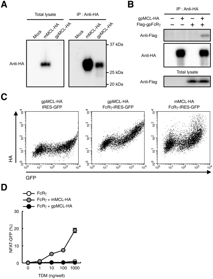
Figure 7. Characterization of gpMCL.
(A) Immunoblot of gpMCL. HEK293T cells were transfected with HA-tagged mMCL or gpMCL. Total lysates were blotted with anti-HA mAb (left panel) or immunoprecipitated with anti-HA pAb and blotted with anti-HA mAb (right panel). (B) gpMCL is associated with gpFcRγ. HEK293T cells were transfected with HA-tagged gpMCL alone or together with Flag-tagged gpFcRγ Total lysates were immunoprecipitated with anti-HA pAb and blotted with anti-Flag pAb and anti-HA mAb. Total lysates were also blotted with anti-Flag pAb. (C) Surface expression of gpMCL. HEK293T cells were transfected with HA-tagged gpMCL alone or together with gpFcRγ-IRES-GFP. HEK293T cells were also transfected with HA-tagged mMCL together with mFcRγ-IRES-GFP. Surface expression of gpMCL or mMCL was detected by anti-HA pAb. (D) gpMCL fails to recognize TDM. Indicated reporter cells were stimulated with plate-coated TDM for 24 h. Induction of NFAT-GFP was analyzed by flow cytometry. Data are presented as mean ± s.d. (D) and representative of two or three separate experiments.

