
Figure 2. Validation of proline-hydroxylated HIF-1α specific antibodies on formalin-fixed paraffin embedded cell pellets.
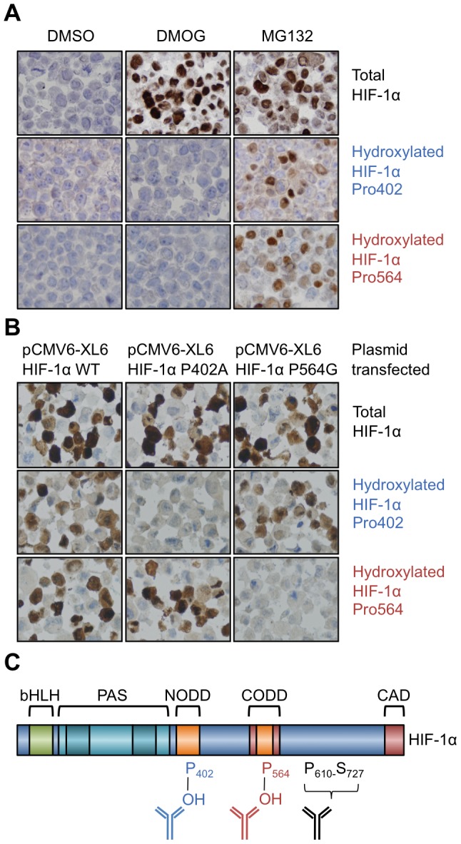
A. MCF7 cells treated with vehicle (DMSO), DMOG or MG132 and probed with the indicated antibodies. B. 786-o cells transfected with either HIF-1α WT, P402A or P564G and probed with the indicated antibodies. C. Domain structure of HIF-1α and antibody binding regions. The figure highlights: basic helix-loop-helix (bHLH) domains; PAS domain; the amino-terminal oxygen-dependent degradation domain (NODD) the carboxy-terminal oxygen-dependent degradation domain (CODD) and the carboxy-terminal activation domain (CAD).