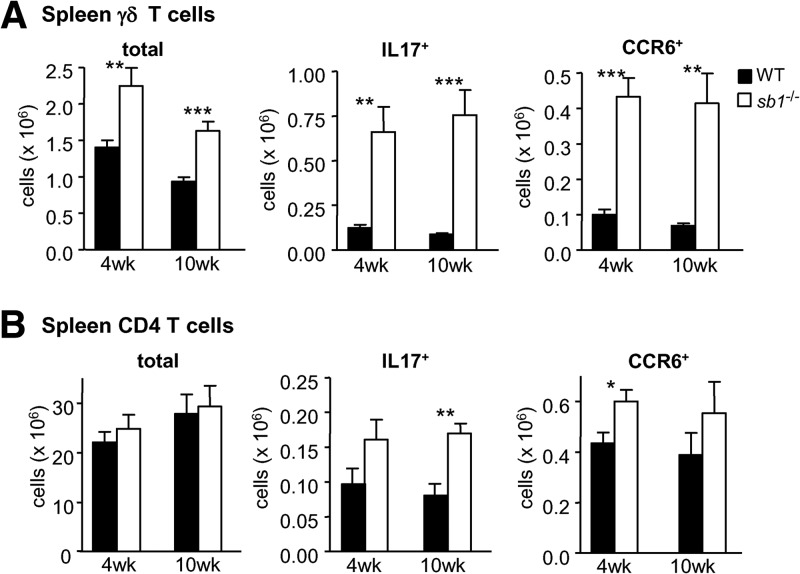
Figure 3. Increase of IL-17+ and CCR6+ γδ T cells in spleens of serpinb1a−/− mice.
Splenocytes of 4- and 10-week-old, naive WT and serpinb1a−/− mice were evaluated for surface antigens or IL-17 expression as in Fig. 1. Cells were gated on lymphocytes (CD45+CD11bneg) and then on γδ TCR or CD4. (A) γδ Cells. Shown are total, IL-17+, and CCR6+ γδ T cells. (B) CD4 cells. Shown are total, IL-17+, and CCR6+ CD4 cells. Means ± sem for eight mice/group at 4 weeks and six mice/group at 10 weeks, each from two experiments. *P < 0.05; **P < 0.01; ***P < 0.001. Related findings are in Supplemental Fig. 2.

