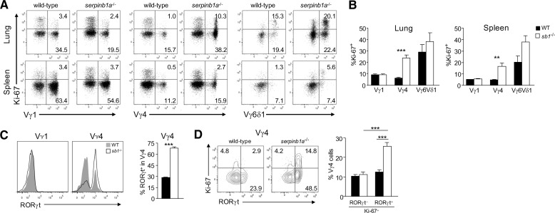
Figure 6. Selectively increased proliferation of serpinb1a−/− Vγ4+ and Vγ6/Vδ1+ γδ T cells.
Freshly isolated lung (A and B) and spleen (A–D) cells of naive WT and serpinb1a−/− mice were surface-stained with γδ subset antibodies, as in Fig. 4, and intracellularly for (A, B, and D) the proliferation marker Ki-67 and (C and D) RORγt. (A) Dot plots showing Ki-67 staining of Vγ1+, Vγ4+, and Vγ6/Vδ1+ subsets. (B) Ki-67+ cells quantified as percentage within each subset. (C) RORγt staining of Vγ1+ and Vγ4+ cells. (Left) Flow cytometry plots. (Right) RORγ+ cells quantified within the Vγ4+ subset. (D) Vγ4 cells costained for Ki-67 and RORγt. (Left) Contour plots. (Right) Quantitation of Ki-67+ RORγneg and Ki-67+ RORγ+ cells within the Vγ4 subsets. Means ± sem or representative data for four mice/group. **P < 0.01; ***P < 0.001.

