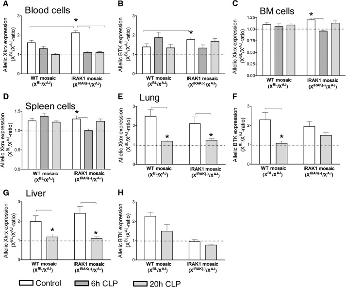
Figure 4. Allelic Xkrx and BTK expression ratios in WT-mosaic and IRAK1-mosaic animals under baseline and septic conditions.
Circulating WBC (A and B), BM cells (C), and splenocytes (D) from control animals (n=8) and mice septic for 6 h (n=8) or 20 h (n=8–12) were analyzed for allelic mRNA expression ratios of Xkrx (A, C, and D) and BTK (B). Lung and liver (n=6) were also analyzed, 20 h after CLP (E–H). Mean ± sem. *Statistically significant difference, as indicated by connecting lines. Dotted line indicates 1/1-ratio.

