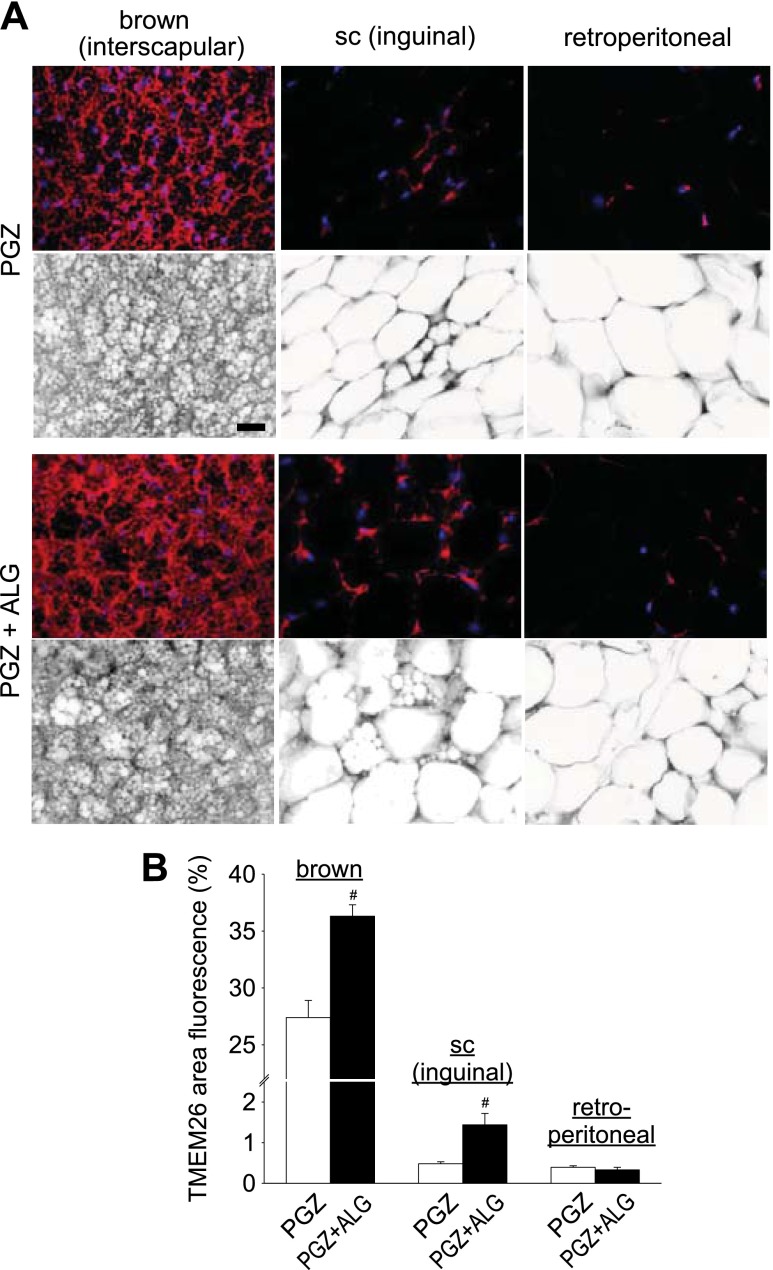
Fig. 6.
ALG enhanced beige cell marker transmembrane protein 26 (TMEM26) expression in interscapular brown adipose tissue (BAT) and inguinal sc WAT in the presence of PGZ. A: immunofluorescene double staining for TMEM26 and DAPI (nuclei marker) of interscapular BAT, inguinal sc WAT, and retroperitoneal WAT in PGZ and PGZ + ALG. Top: images under fluorescence mode; bottom: images under inverted mode. Each pair image (top and bottom) is the same area. Scale bar, 100 μm. B: TMEM26-positive area (%) of interscapular BAT, inguinal sc WAT, and retroperitoneal WAT in PGZ and PGZ + ALG. #P < 0.05 vs. PGZ alone; n = 4–5/group.

