
FIGURE 1.
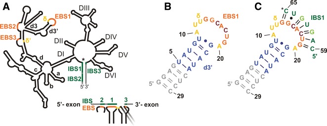
Secondary structures of a group IIB intron and the constructs under investigation. (A) Schematic representation of a group II intron with its six domains (labeled DI–VI) arranged around a central wheel. In DI, the four main branches a–d are labeled; the d3′ hairpin containing EBS1 is a substructure of branch d. The intron is shown in black, and the 3′ and 5′ exons are shown in gray. (*) The 5′ splice site. (Orange) EBS sequences; (green) IBS sequences; (yellow) the two nucleotides (nt) forming the δ–δ′ base pair. Below, the base-pairing scheme of the three EBS•IBS contacts and the δ–δ′ contact is shown. The latter is required to ensure that the 5′ and 3′ exons are bound close to each other in the active site of the intron (see also text). Secondary structures of d3′EBS1 (B) and d3′EBS1•IBS1 (C). (*) The location of the 5′ splice site. (Gray) Additional nucleotides added for better transcription yields; (blue) the helical stem of the native d3′ sequence; (yellow) the unpaired nucleotides; (orange) EBS1 (nucleotides G13–G19); (green) IBS1 (C59–C65). Light green letters in IBS1 and dark red letters in EBS1 mark mutations introduced for the sake of stability (see text and Kruschel and Sigel 2008).