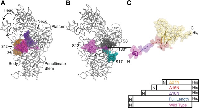
FIGURE 1.

Structural relationship of S12 to other SSU components. (A) Positions of S12 (magenta), S4 (orange), and S5 (violet) in the three-dimensional structure of the E. coli 30S subunit. 16S rRNA is gray and other r-proteins omitted for clarity. All figures containing three-dimensional structures were prepared using PyMOL and Protein Data Bank file 2AW7 (Schuwirth et al. 2005). (B) Location of S12 (magenta), S8 (black), and S17 (teal) in the three-dimensional structure of the 30S subunit. 16S rRNA is gray and other r-proteins omitted for clarity. (C) Three-dimensional structure of S12 as found in the 30S subunit, rotated 180° with respect to B, with the variants color coded. S12WT is magenta, S12Full Length is blue, S12▵10N is purple, S12▵15N is red, and S12▵27N is orange.
