
Figure 3. Characterization of oxTrp72-apoA1 recovered from human atherosclerotic plaque.
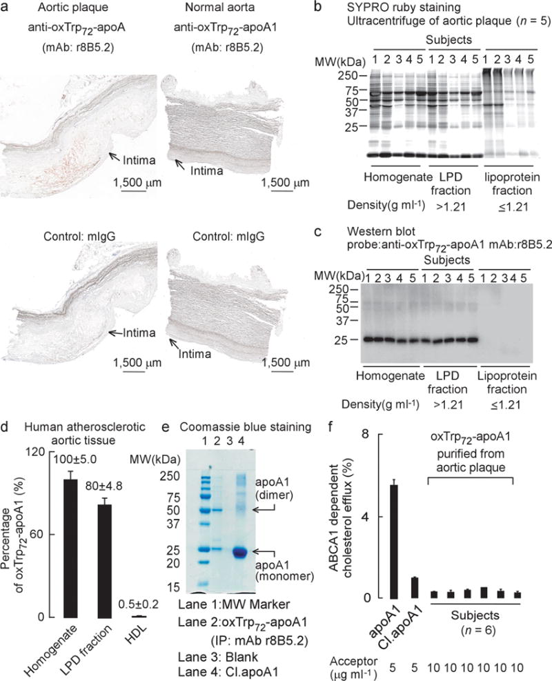
(a) Immunohistochemical staining of fresh frozen normal and atherosclerotic sections of human aorta. OxTrp72-apoA1 was detected with mAb r8B5.2. Mouse IgG was used as control. Positive staining: Red; Blue, haematoxylin counter stain. (b) SDS-PAGE, protein (SYPRO ruby) staining and (c) Western blot of human aortic plaque homogenate and the indicated fractions separated by buoyant density ultracentrifugation. (d) Level of oxTrp72-apoA1 (as a percentage relative to total apoA1) recovered in human aortic tissue homogenate vs. the lipoprotein (d ≤ 1.21 g/ml) and lipoprotein depletion (LPD) fractions (d > 1.21 g/ml). N.A. = non-detectable. Means ± S.D. of triplicate determinations are shown. (e) SDS-PAGE and Coomassie blue (protein) staining of mAb r8B5.2 immunopurified oxTrp72-apoA1 from human aortic tissue (lane 2). As positive control, apoA1 exposed to the MPO/H2O2/Cl− system (at 10:1 molar ratio of H2O2 to apoA1), Cl.apoA1, was loaded in lane 4. ApoA1 monomer and dimer (MS sequence confirmed) are indicated. (f) ABCA1-dependent cholesterol efflux activity of oxTrp72-apoA1 immunoaffinity purified from individual human aortic plaque. Native apoA1 and Cl.apoA1 served as controls.