
Figure 4. Characterization of oxTrp72-apoA1 recovered from human plasma.
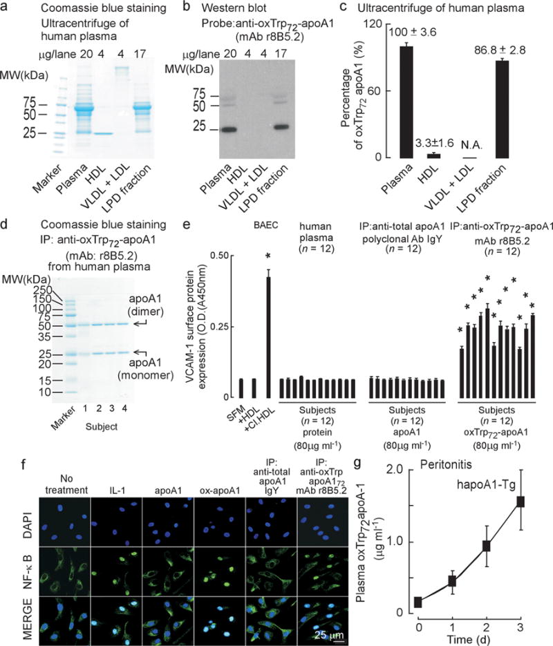
(a) SDS-PAGE, Coomassie blue staining and (b) oxTrp72-apoA1 (mAb 8B5.2) western blot of human plasma proteins before and after buoyant density ultracentrifugation separation into the indicated fractions. (c) Distribution of oxTrp72-apoA1 in plasma. Results shown are mean ± S.D. of triplicate determinations of four pooled healthy donor plasma pools. (d) SDS-PAGE with Coomassie blue staining of immuno-purified (using mAb r8B5.2) oxTrp72-apoA1 from plasma from four donors. (e) Bovine aortic endothelial cell (BAEC) were incubated with aliquots of individual subjects’ plasma, total apoA1 (recovered using immobilized anti-total apoA1 IgY) or oxTrp72-apoA1 (using immobilized mAb r8B5.2) and surface VCAM-1 protein expression was quantified by cell-based ELISA as described under Methods. Serum free media (SFM), HDL, or HDL previously exposed to the MPO/H2O2/Cl− system (Cl.HDL, at H2O2:apoA1, 10:1, mol:mol) were used as vehicle, negative and positive controls, respectively. *, P < 0.05. (f) Nuclear translocation of NF-κB: Confocal immunofluorescent analysis of BAEC cells, untreated or treated with the following: IL-1 (positive control) native (apoA1) or MPO-oxidized human apoA1 (ox-apoA1), plasma-derived total apoA1 immunopurified using IgY to apoA1; plasma oxTrp72-apoA1 immuno-purified using r8B5.2. NF-κB p65 (E498) staining is green and nuclear counterstaining (DAPI) is blue. (g) Human apoA1-Tg mice were injected i.p. with zymosan (T=0). Plasma oxTrp72-apoA1 levels were quantified at the indicated times by sandwich ELISA using mAb r8B5.2 as capture antibody, and mAb 10G1.5 (anti-total apoA1) as detecting antibody, as described in Methods.