
Figure 6. Formation of a dysfunctional apoA1 form, oxTrp72-apoA1, within human atherosclerotic lesions.
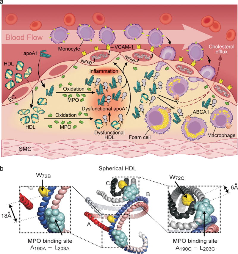
(a) Schematic summary illustrating formation of an abundant dysfunctional apoA1 form in human atheroma generated by MPO. (b) Model of spherical HDL demonstrating close spatial proximity between MPO binding site on one apoA1 chain, and Trp72 on the contralateral anti-parallel apoA1 chain. Shown are the longest (18Å) and shortest (6Å) predicted distances among two of the three apoA1 chains43. The apoA1 polypeptide chains are depicted in red and slate gray. W72, yellow; MPO interaction site with apoA1, light blue.