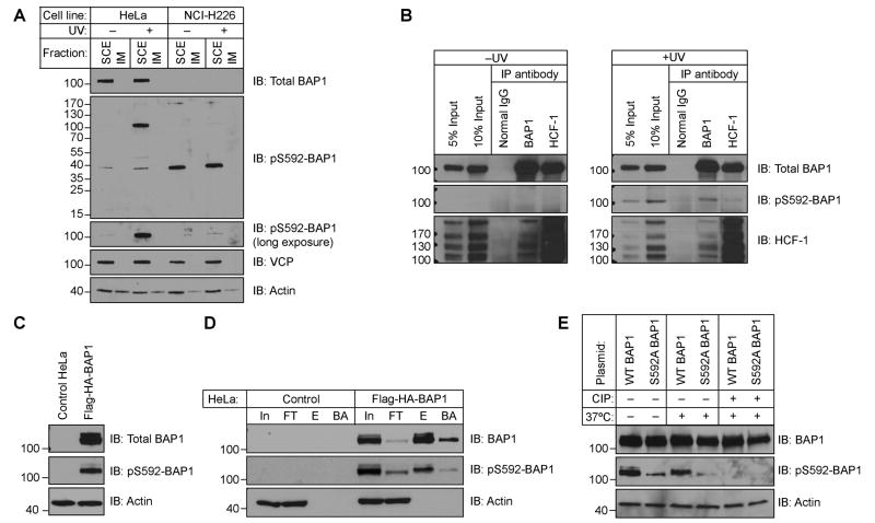
Figure 1.
BAP1 is phosphorylated at S592 following UV irradiation. (A) The pS592-BAP1 antibody recognizes a ∼100 kDa band after UV-treatment in HeLa cells but not in the BAP1-null NCI-H226 cells. Cells were mock/UV-treated with 1 mJ/cm2 and allowed to recover 3 h before analyzing soluble cell extract (SCE) and insoluble material (IM). (B) IP of endogenous BAP1 and HCF-1 retrieves pS592-BAP1. Cells were mock- or UV irradiated with a 50 mJ/cm2 dose and allowed to recover 2 h before IP from SCEs. (C) Overexpressed Flag-HA-BAP1 is phosphorylated in the absence of UV. (D) The pS592-BAP1 signal is immunoprecipitated with anti-Flag agarose (In, Input; FT, Flow thru; E, eluate; BA, boiled agarose). (E) Phospho-specificity of the pS592-BAP1 antibody.

