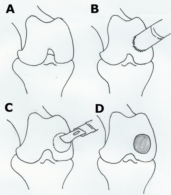
Figure 1.
Surgical procedures for cartilage defect. (A) The joint was exposed after anesthesia. (B) trephine (5 mm in diameter) was used to create a cartilage defect with a round shape on the femoral medial condyle. (C) Cartilage within the circle was removed with a knife gently. (D) The subchondral bone was exposed as a severe chondral injury.

