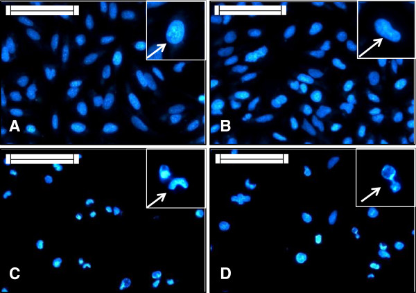
Figure 3.
HeLa cells grown on sterile cover slips were treated with IC50 doses of the AMEs for 24 hours. Then they were stained with Hoechst 33258 to study the nuclear morphology. Scale indicates 50 μm. Inset shows magnified view. (A) Non-treated cells, the arrow shows normal looking nucleus; (B) Vehicle control cells, the arrow shows normal elliptical nucleus; (C) E. intestinalis extract treated sets, the arrow shows nuclei with deformity; (D) R. riparium treated cells, the arrow shows highly condensed nuclei.

