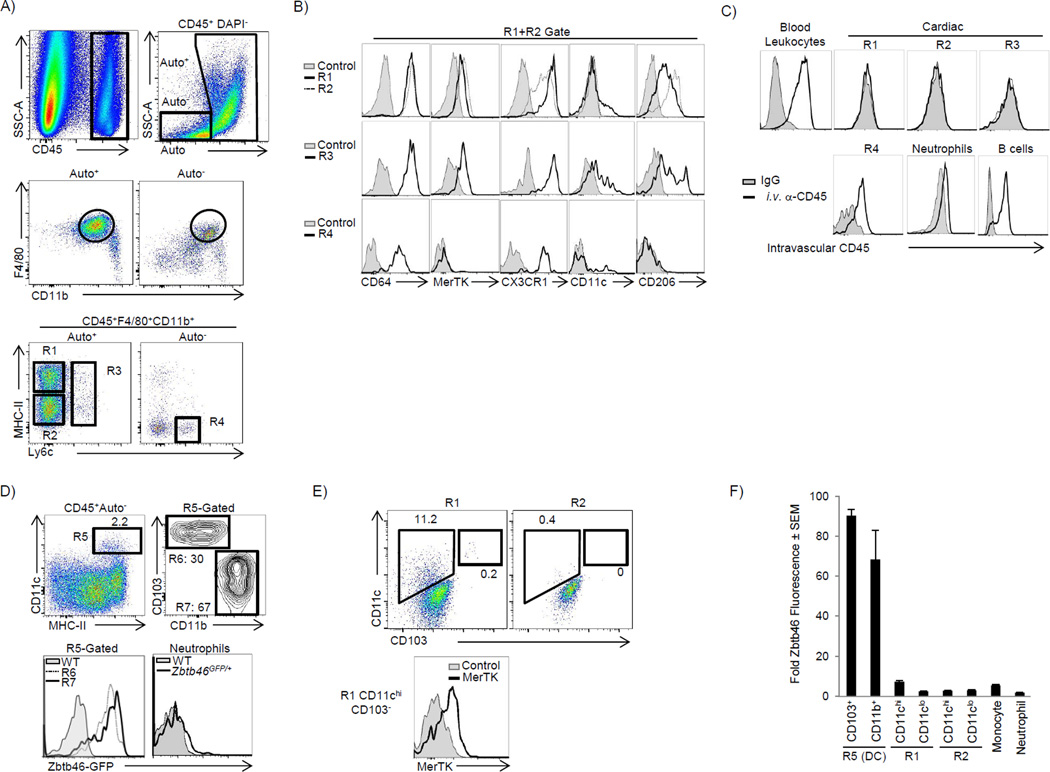
Figure 1. The adult heart contains distinct cardiac macrophage subsets (see also Fig S1).
Cardiac single cell suspensions were analyzed by flow cytometry – see full gating strategy in Fig S1A. A) CD45+ leukocytes were identified, doublets excluded (by FSC-W vs. FSC-A) and dead cells excluded by DAPI. Live cells were stratified by autofluorescence (Auto+ or Auto−), gated on F4/80+ CD11b+ myeloid cells and further stratified by MHC-II and Ly6c expression. R1: MHC-IIhi macrophages. R2: MHC-IIlo macrophages. R3: Ly6c+ macrophages. R4: Ly6chi monocytes. B) Cardiac samples were labeled with isotype control antibody (Control) or with the indicated antibodies. Expression of CX3CR1 was assessed in Cx3cr1GFP/+ mice and compared with WT mice (Control). C) To label intravascular leukocytes, mice were injected i.v. with anti-CD45 and sacrificed 5 min later. Cells were gated as in panel A and intravascular CD45 fluorescence is shown. B cells were B220+ MHCII+ CD11b−F4/80− and neutrophils were Ly6g+ CD11b+ F4/80−. D) The Auto− subset contained the majority of cardiac DCs (Total DCs - R5), which were made up of CD103+ CD11b− (R6) and CD103−CD11bHi (R7) DCs. ZBTB46 expression was assessed in Zbtb46GFP/+ mice in R6 and R7, and compared to Ly6g+ neutrophils and compared to WT mice. E) Expression of CD11c and CD103 within the primary macrophage gates (R1 and R2). F) Relative Zbtb46 fluorescence ratio in myeloid subsets within the myocardium. The geometric mean fluorescence intensity (gMFI) in each subset in Zbtb46GFP/+ mice was divided by gMFI of that subset in WT mice. The primary macrophages populations in R1 and R2 were further stratified by CD11c expression. N=4–8.

