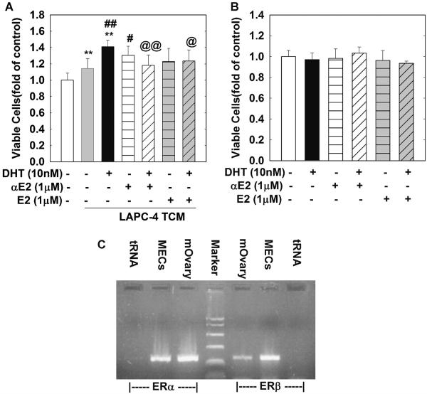
Fig. 3.
Co-administration of αE2 or βE2 with DHT in LAPC-4 cells inhibits DHT enhancement of TCM-induced MEC cell proliferation. In (A) MECs were plated in 96-well plates and treated for 48 hr with TCMs collected from LAPC-4 cells treated with a vehicle control, or αE2 (1 μM), or βE2 (1 μM) in the presence or absence of DHT as indicated. In (B) MECs were plated in 96-well plates and treated with vehicle control, DHT (10 nM), αE2 (1 μM), or βE2 (1 μM) alone or in combination for 48 hr. (C) is a representative RT-PCR analysis of mouse ERα and ERβ expression in MECs. Mouse ovary (mOvary) and tRNA were used as positive and negative control, respectively. The data are the means ± SEM SEM of three independent triplicate experiments. **P < 0.01 compared to vehicle control; ##P < 0.01 and #P < 0.05 compared to TCM-vehiclegroup; and @@P < 0.01 and @P < 0.05 compared to TCM-DHT10 nM group.

