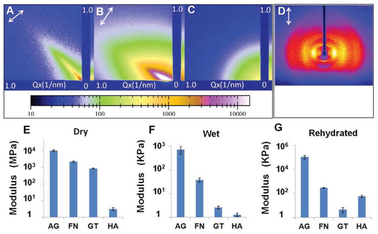
Fig. 3.
X-ray scattering diffraction patterns and tensile moduli of hydrogel fibres in dry and wet states. (A–B) Small angle X-ray scattering (SAXS) patterns of the dry (A) and wet (B) calcium alginate hydrogel fibres confirming an alignment axis along the microfibre orientation indicated by the arrows. (C) SAXS pattern of alginate hydrogel prepared by hand extrusion suggesting an isotropic structure. (D) Wide angle x-ray scattering pattern of the dry alginate microfibres confirming the polymer chain alignment along the fibre axis as indicated by the arrow. (E–G) Tensile moduli of alginate (AG), fibrin (FN), gelatin (GT) and hyaluronic acid (HA) hydrogel fibres in dry (E), wet (F) and rehydrated form (G).

