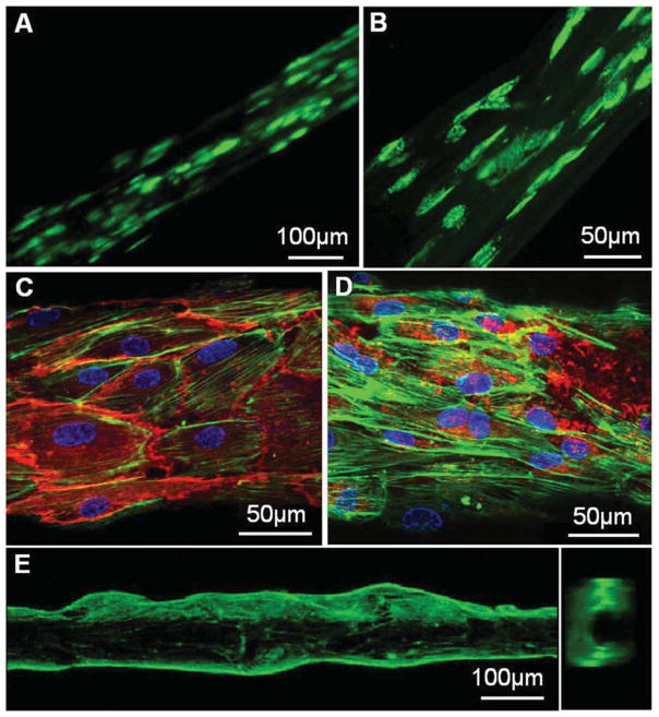
Fig. 4.
Fluorescence micrographs of cells grown inside or outside fibrin hydrogel microfibres. (A–B) ASCs with CellTracker™ green CMFDA dye were encapsulated in 0.75 wt% fibrin hydrogel fibres. After 2 days (a) and 5 days (b) of culture, cells spread out showing alignment along the fibre axis. (C–D) F-actin filament (green) staining of human ECFCs cultured on the surface of a rehydrated fibrin microfibre, overlapped with immunofluorescence staining for CD31 (red) (C) or vWF (red) (D) and DAPI (blue) staining for cell nuclei. (E) A side view and a cross-section view of the sample shown in (C–D), indicating that the differentiated ECFCs grew into a confluent tubular structure.

