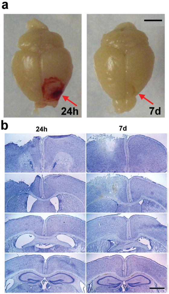
Figure 1.

Frontal TBI induces a distinct unilateral lesion. Panel (a) depicts representative photographs of whole perfused brains collected at 24 h and 7 d post-injury (scale bar = 3 mm). Panel (b) illustrates representative coronal sections stained with cresyl violet to delineate tissue damage at 24 h and 7 d. Images were captured at ~Bregma +1.70, +0.50, -0.10 and -0.15 mm respectively, from top to bottom.
