Figure 2.

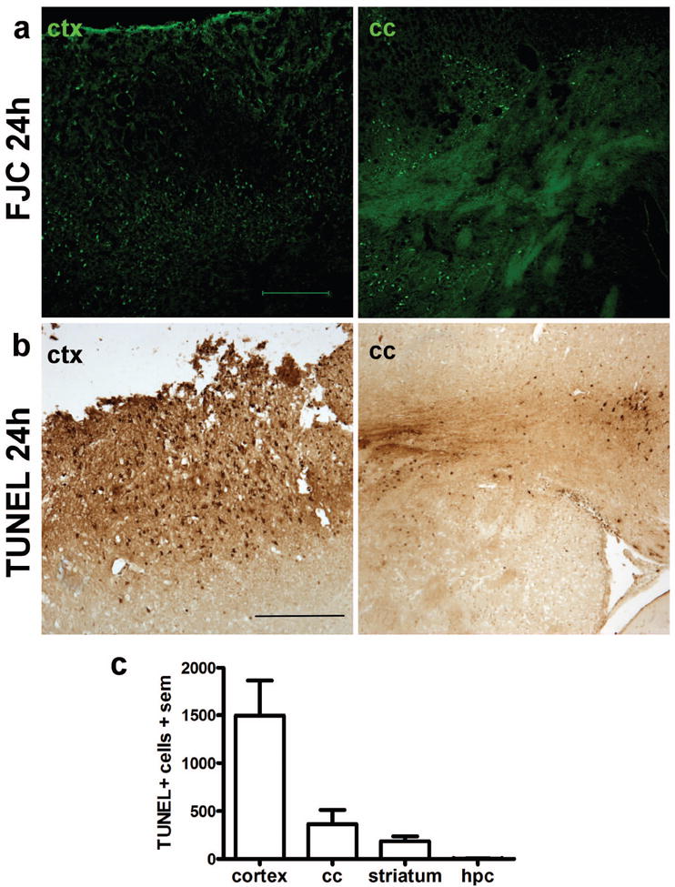
Local cell death in the dorsal cortex and subcortical structures after frontal TBI. Degenerating neurons were labeled with Fluoro-Jade C at 24 h post-injury (a), revealing abundant neuronal damage in the peri-contusional cortex and dorsal striatum (scale bar = 200 μm). Cell death was confirmed by TUNEL staining (b), which was widespread in the peri-contusional cortex as well as in the underlying corpus callosum and striatum (scale bar = 250 μm). TUNEL-positive cells were summed across 5 sections per brain at 24 h post-injury (c), highlighting the regional localization of acute cell death after frontal TBI (one-way ANOVA, F3,28=11.07, p<0.0001; n=8).
