
Figure 3.
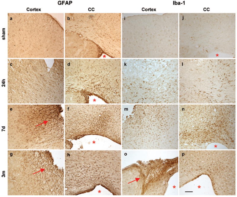
Immuno-reactivity for astrocytes (GFAP) and reactive microglia/macrophages (Iba-1) is evident in the ipsilateral dorsal cortex (c, e, g, k, m, o) and corpus callosum (d, f, h, j, l, n, p) after frontal TBI. Sham operation did not induce gliosis at 24 h (1st row). At 24 h post-injury (2nd row), only few reactive astrocytes are noted, although Iba-1 reactivity was increased above sham levels. By 7 d (3rd row), both GFAP+ astrocytes and Iba-1+ reactive microglia were visibly accumulating in the peri-contusional parenchyma. By 3 months post-injury, GFAP+ astrocytes formed a glial scar at the cortical lesion edge. The lesion core (o) also stains strongly for Iba-1 at these times. Scale bar =100 μm. Arrows indicate a glial scar; asterisks highlight the enlarged lateral ventricle.