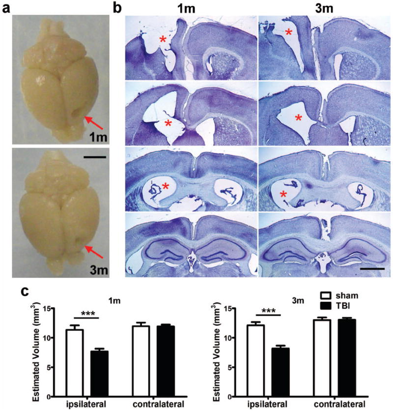
Figure 5.

Frontal TBI results in cavity formation and a volumetric reduction in the injured cortex. At 1 and 3 months post-injury, a pronounced unilateral cortical cavity was evident in whole perfused brains (arrows in panel a, scale bar = 3 mm) and representative dorsal cortex sections stained with cresyl violet (b, scale bar = 500 μm). Images were captured at ~Bregma +1.70, +0.50, -0.10 and -0.15 mm respectively, from top to bottom for each time point shown. Note the enlarged lateral ventricle ipsilateral to the injury site at 1 and 3 months (asterisks). Remaining dorsal cortex was quantified at both 1 and 3 months post-injury (c), demonstrating significant volumetric loss in the ipsilateral dorsal cortex at both time points (2-way ANOVA post-hoc, p<0.0001; n=10/group).
