
Figure 1.
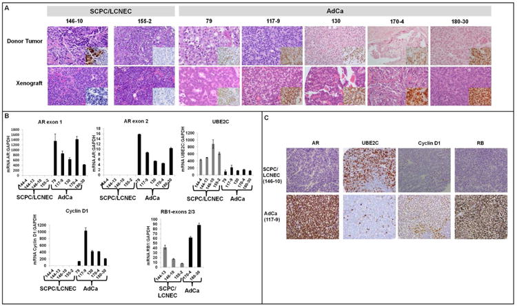
A, Representative images of the xenografts and their donor tumors, with hematoxylin and eosin staining; the insets show IHC staining for AR. Note that the MDA PCa 146-10 donor tumor was mixed, containing adenocarcinoma (AdCa) and SCPC components. The inset shows that the AdCa component was AR+, whereas the SCPC component was AR–. (Chr: chromogranin; Syn: synaptophysin. Original magnification of all images except 155-2, ×200, original magnification of 155-2 images, X100). B, mRNA levels of AR (exons 1 and 2), UBE2C, cyclin D1, and RB1 exons 2/3 in SCPC/LCNEC (gray bars, MDA PCa 144-4, 144-13, 146-10, and 155-2) and AdCa xenografts (black bars, MDA PCa 79, 117-9, 130, 170-4, and 180-30). C, Representative images of AR, UBE2C, cyclin D1, and RB immunostaining in SCPC/LCNEC and AdCa xenografts. Original magnification, ×200.