Figure 3.
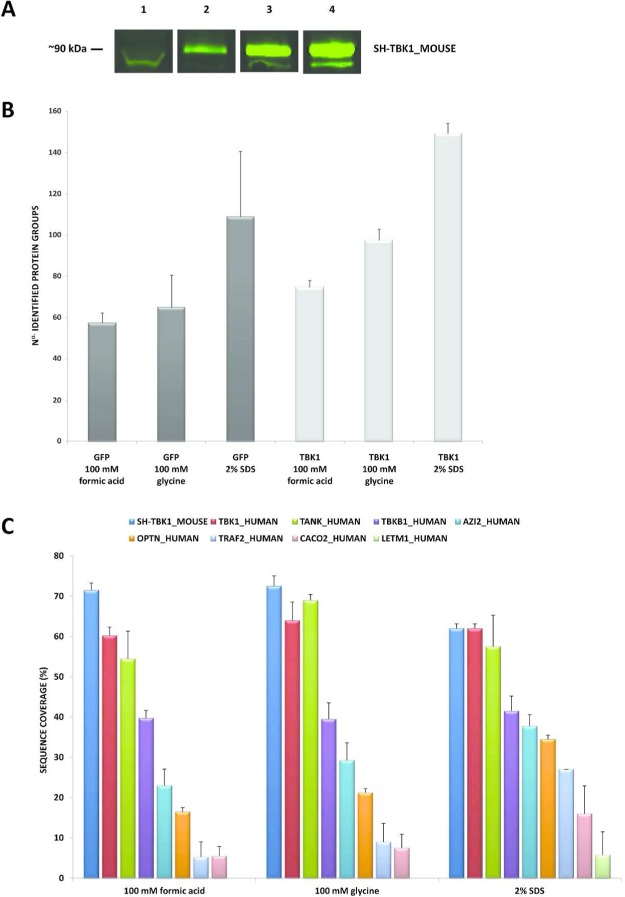
(A) Fluorescent anti-HA immunoblot of the second step eluates from the anti-HA agarose beads for SH-TBK1_MOUSE. Elutions were performed with 100 mM formic acid (lane 2), 100 mM glycine (lane 3), and 2% SDS (lane 4). 100 μg whole cell extract (lane 1); lanes 2–4, 1% v/v loaded. Immunoblotting was performed with mouse HA.11 antibody (1:3000) followed by goat antimouse antibody (800 nm) (1:15 000). (B) Total number of protein groups identified for the SH-GFP and SH-TBK1_MOUSE purified protein complexes for the three elution conditions: formic acid, glycine, and 2% SDS (n = 4). (C) Average protein sequence coverage (%) for the purified protein complexes from the three elution conditions: formic acid, glycine, and 2% SDS (n = 4). Data presented are for SH-TBK1_MOUSE; the core protein complex interactors, TBK1_HUMAN, TANK_HUMAN, TBKB1_HUMAN, AZI2_HUMAN; plus the additional interactors OPTN_HUMAN, TRAF2_HUMAN, CACO2_HUMAN, and LETM1_HUMAN. GFP, green fluorescent protein; TBK1, TANK binding kinase 1; HA, hemagglutinin; SDS, sodium dodecylsulphate.

