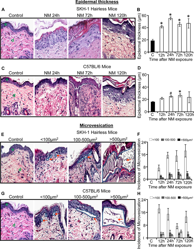
Fig. 1.
Topical application of NM on to the dorsal skin of SKH-1 hairless and C57BL/6 mice causes an increase in epidermal thickness and microvesication. Mice were exposed topically to either 200 µL of acetone alone or with NM (3.2 mg) in 200 µL acetone for 12–120 h and skin tissue was collected, processed, sectioned and subjected to H&E staining as detailed under materials and methods. The H&E stained skin tissue sections from both the mouse strains were analyzed for epidermal thickness (A–D) and microvesication (E–H). The NM-induced epidermal thickness in SKH-1 hairless and C57BL/6 mice is shown in representative pictures (A and C), and was further quantified (B and D) as detailed under materials and methods. NM-induced microvesication in SKH-1 hairless and C57BL/6 mice is shown in representative pictures (E and G), which was further quantified according to their sizes (<100µm2, 100–500 µm2, >500 µm2; F and H) as detailed under materials and methods. Data presented are mean ± SEM of three-five animals in each treatment group. Statistical significance of difference between NM and control groups were determined by one way ANOVA followed by Bonferroni t-test for pair wise multiple comparisons. *, p<0.001; $, p<0.01 and #, p<0.05 as compared to control. e, epidermis; d, dermis. Red arrows show the different sizes of microvesication.

