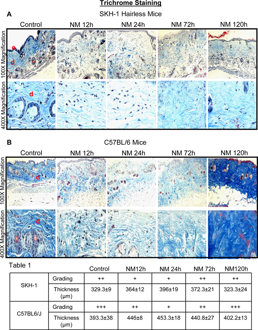
Fig. 3.
Topical application of NM causes changes in dermal collagen staining and thickness in SKH-1 hairless and C57BL/6 mice skin. Mice were exposed topically to either 200 µL of acetone alone or with NM (3.2 mg) in 200 µL acetone for 12–120 h and skin tissue was collected, processed, sectioned and subjected to Gomori’s trichrome staining. NM-induced changes in collagen staining and organization, and dermal thickness in Gomori’s trichrome stained skin sections from SKH-1 hairless and C57BL/6 mice is shown in representative pictures (A and C), staining was further graded (+, weak staining; ++ strong staining; +++, very strong staining) and dermal thickness quantified (Table 1) as detailed under materials and methods. Collagen stained royal blue and nuclei stained dark blue/black. Grading and thickness were observed in three-five animals at each time point after NM exposure. e, epidermis; d, dermis

