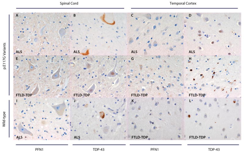
Figure 2. PFN1 expression in patients with and without p.E117G variants.
Spinal cord motor neurons of the two patients with p.E117G variants lack PFN1 inclusions (A, E). Temporal cortex for these two patients does not demonstrate neuronal PFN1 inclusions either (C, G). Two patients without the p.E117G variant, one of whom was diagnosed with ALS (I) and one with FTLD-TDP (K), who were photographed as controls for this variant, display the same pattern as patients with p.E117G variants in the spinal cord and temporal cortex, respectively. For the two patients with p.E117G variants, TDP-43 staining is shown both in the spinal cord (B, F) and temporal cortex (D, H): the ALS patient demonstrates TDP-43 positive inclusions in the spinal cord (TDP-43 type 3, B), whereas the FTLD-TDP patient shows TDP-43 positive inclusions in the temporal cortex (TDP-43 type 2, H). TDP-43 positive inclusions are also present in the spinal cord of the wild-type ALS patient (TDP-43 type 3, J), and in the temporal cortex of the wild-type FTLD-TPD patient (TDP-43 type 1, L). Magnification: 40×.

